Rocha Twins Doing Fine,
First Fotos Released
Doctors at Lucile
Packard Children's
Hospital at Stanford
said Wednesday formerly
conjoined Costa Rican
twins Yurelia and
Fiorella Rocha-Arias are
continuing to recover
from their Nov. 12
separation surgery.
Yurelia was released
from the hospital’s
cardiovascular intensive
care unit on
Tuesday, Fiorella is
expected to join her by
the end of the week,
according to doctors.
"Everyone’s thrilled
that they‘re progressing
so well," said Gail
Wright, MD,
cardiovascular intensive
care physician. "Moving
out of intensive care is
a very big step."
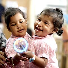
Doctors also released
the first post-surgery
photo of the twins. It
shows one child in a
hospital bed with the
girl's mother holding
the second child nearby.
Doctors said Fiorella's
recovery lags slightly
behind her sister’s
because she returned to
surgery on Nov. 19 for a
brief modification of
her chest reconstruction
and repair of her skin
closure.
She also needs a couple
more days for her
respiratory issues to
completely clear.
Yurelia's last surgery
happened on Nov. 14 when
her life-threatening
congenital anomaly,
known as double outlet
right ventricle, was
corrected by Frank
Hanley, chief of
pediatric cardiothoracic
surgery.
The girls were connected
at parts of their hearts
and shared a liver
before their successful
nine-hour separation
surgery earlier this
month.
"It went really well,"
said cardiothoracic
surgeon Frank Hanley, of
the six-hour correction
and reconstruction.
Hanley is the hospital’s
chief of pediatric
cardiothoracic surgery
and an internationally
known specialist in
treating complex
congenital heart defects
in children, Dicks said.
DORV occurs early in
pregnancy when the heart
is developing, Dicks
said.
In children with DORV,
both the pulmonary
artery and the aorta are
connected to the right
ventricle.
In a normal heart, the
aorta is connected to
the left ventricle and
delivers oxygenated
blood throughout the
body.
Children with DORV have
trouble getting enough
oxygen to their tissues
and their hearts must
work abnormally hard to
support life, Dicks
said.
Surgeon Created Tunnel
In Girl's Heart
For the separation
surgery, Hanley created
a tunnel from the left
ventricle through the
right ventricle to the
aorta to correct the
flow of blood in
Yurelia’s body, Dicks
said.
The tunnel passed
through and sealed a
hole between the sides
of Yurelia’s heart that
had allowed mixing of
oxygen-rich and
oxygen-poor blood, Dicks
said.
Hanley also widened an
area in Yurelia’s heart
near the pulmonary valve
that had been
constricting the flow of
blood to Yurelia’s
lungs, Dicks said.
Because Yurelia and
Fiorella were joined at
the chest and abdomen
and their hearts were
connected, it was
impossible to correct
Yurelia’s DORV before
the twins were
separated.
"These are two very
strong little girls,"
lead surgeon Gary
Hartman said.
Their mom, Maria, and
dad, Jose Luis, have
remained at the girls'
bedside for weeks, Dick
said |
|